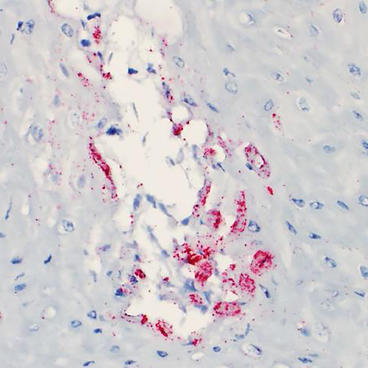
Senecavirus A - SVA (Seneca Valley Virus) belongs to Genus Senecavirus within the Picornaviridae family. The clinical signs are characterized by vesicles/blister and coalescing erosions on the snouts and coronary bands in clinically infected sows, nursery, and finishing pigs (Figure 1). Increased mortality in neonatal pigs has also been sporadically reported. The picture to the right is a microvesicle in the epidermis (skin) of a pig infected with SVA. SVA mRNA is stained by the red dots. (Provided by Dr. F. Vannucci at the MNVDL). It is imperative to differentiate SVA from other viruses that cause skin vesicles/blisters such as the Foot and Mouth Disease virus. If you see vesicles/blisters on your pigs, call your veterinarian immediately.
Archived resources on Senecavirus A
- Senecavirus A in swine powerpoint presentation (PDF)
- UMN VDL experience with Seneca Valley Virus (PDF)
- VDL Develops Antibody Test for SVV (article) (PDF)
- Journal Article on Seneca Valley Virus (PDF)
- Information from AASV on Seneca Valley Virus (PDF)
- USDA Recommendations for Swine with Potential Vesicular Disease (PDF)
- Longitudinal Study of Senecavirus A (PDF)